
Introduction
Fatty liver is a common liver condition in which excess fat accumulates inside liver cells. It may not cause noticeable symptoms in the early stages, which is why it is sometimes discovered during a routine abdominal ultrasound or blood test.
Two tests commonly discussed during fatty liver evaluation are abdominal ultrasound and FibroScan. Although both use ultrasound-based technology, they provide different information about the liver.
The main difference is that an abdominal ultrasound creates images of the liver and other abdominal organs, whereas FibroScan measures liver stiffness and estimates liver fat using specific measurements called LSM and CAP.
Understanding the difference between ultrasound and FibroScan can help you discuss the right investigations with your gastroenterologist.
1. What Is an Abdominal Ultrasound and How Does It Detect Fatty Liver?
An abdominal ultrasound, also known as USG abdomen, is a non-invasive imaging test that uses high-frequency sound waves to create images of internal organs. It is commonly used to examine the liver, gallbladder, kidneys, pancreas and other abdominal structures.
How Does an Abdominal Ultrasound Work?
During the examination, a sonographer or radiologist applies a gel to the abdomen and moves a handheld device called a transducer over the skin.
The transducer sends sound waves into the body and receives the returning echoes. A computer processes these signals to produce images of the abdominal organs on a monitor.
The procedure is generally painless and does not use ionising radiation.
How Does Ultrasound Identify Fatty Liver?
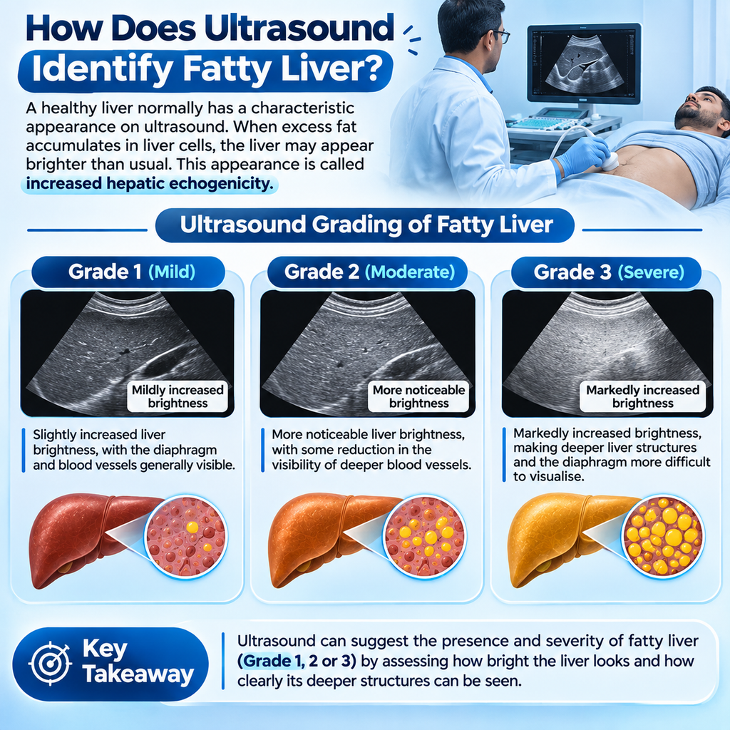
A healthy liver normally has a characteristic appearance on ultrasound. When excess fat accumulates in liver cells, the liver may appear brighter than usual. This appearance is called increased hepatic echogenicity.
Based on the appearance of the liver, radiologists may describe fatty liver as:
- Grade 1 (Mild): Slightly increased liver brightness, with the diaphragm and blood vessels generally visible.
- Grade 2 (Moderate): More noticeable liver brightness, with some reduction in the visibility of deeper blood vessels.
- Grade 3 (Severe): Markedly increased brightness, making deeper liver structures and the diaphragm more difficult to visualise.
These grades provide a visual estimate of fatty changes. They do not directly measure the exact amount of liver fat or determine the stage of liver fibrosis.
What Are the Limitations of an Abdominal Ultrasound?
Although abdominal ultrasound is useful for examining liver structure, it has certain limitations:
- May Miss Mild Fatty Liver: Conventional ultrasound is less sensitive when fat accumulation is mild.
- Cannot Directly Measure Liver Stiffness: It cannot reliably determine the extent of liver fibrosis.
- Does Not Quantify Liver Fat Precisely: The grading is based on visual assessment rather than an exact fat measurement.
- Results Depend on Image Quality: Body habitus, equipment and operator experience can influence image quality and interpretation.
A normal ultrasound does not completely rule out fatty liver, inflammation or liver fibrosis. Further evaluation may be appropriate depending on a person’s medical history and risk factors.
2. What Is a FibroScan Test and How Does It Work?
FibroScan, also known as vibration-controlled transient elastography (VCTE), is a specialised non-invasive test used to assess liver stiffness and estimate liver fat.
Unlike a conventional abdominal ultrasound, which produces images of abdominal organs, FibroScan provides numerical measurements that help doctors assess liver health.
How Is a FibroScan Test Performed?
During the test, the patient usually lies on an examination bed with the right arm positioned above or behind the head.
A healthcare professional places a specialised probe between the lower right ribs over the liver. The probe generates a gentle vibration that produces a shear wave through the liver tissue.
The device measures how quickly the wave travels through the liver. The results help estimate liver stiffness.
The examination is generally painless and does not require needles or surgical incisions.
What Are the Two Main FibroScan Measurements?
FibroScan provides two important measurements:
1. Liver Stiffness Measurement (LSM)
- Measured in kilopascals (kPa).
- Estimates how stiff the liver tissue is.
- Higher stiffness measurements may indicate an increased likelihood of liver fibrosis.
- Results must be interpreted alongside clinical findings and other investigations.
2. Controlled Attenuation Parameter (CAP Score)
- Measured in decibels per metre (dB/m).
- Estimates the amount of fat-related attenuation in the liver.
- Helps assess the degree of hepatic steatosis.
- Does not provide an exact percentage of liver cells containing fat.
The CAP score and LSM provide different types of information. Neither measurement should be interpreted in isolation or treated as a definitive diagnosis.
3. Ultrasound vs FibroScan: What Is the Difference?
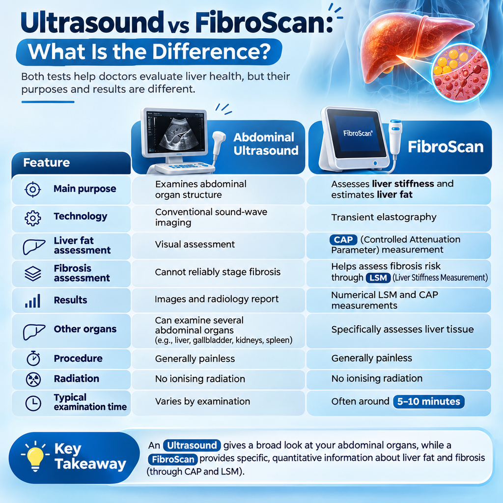
Both tests help doctors evaluate liver health, but their purposes and results are different.
| Feature | Abdominal Ultrasound | FibroScan |
| Main purpose | Examines abdominal organ structure | Assesses liver stiffness and estimates liver fat |
| Technology | Conventional sound-wave imaging | Transient elastography |
| Liver fat assessment | Visual assessment | CAP measurement |
| Fibrosis assessment | Cannot reliably stage fibrosis | Helps assess fibrosis risk through LSM |
| Results | Images and radiology report | Numerical LSM and CAP measurements |
| Other organs | Can examine several abdominal organs | Specifically assesses liver tissue |
| Procedure | Generally painless | Generally painless |
| Radiation | No ionising radiation | No ionising radiation |
| Typical examination time | Varies by examination | Often around 5–10 minutes |
Which test is better for fatty liver?
Neither test replaces the other in every situation. An abdominal ultrasound helps examine liver appearance and other abdominal organs, while FibroScan provides additional information about liver stiffness and estimated fat content.
Your doctor may recommend one or both tests depending on your symptoms, blood test results, medical history and risk factors.
4. Why Is Liver Stiffness Important in Fatty Liver Disease?
Fat accumulation is not the only concern in fatty liver disease. Some people may also develop inflammation and fibrosis, which can affect liver function over time.
Understanding these changes is important because the amount of fat in the liver does not always reflect the extent of liver damage.
Understanding the Stages of Fatty Liver Disease
The progression of fatty liver disease can involve different changes:
1. Steatosis (Fat Accumulation)
Excess fat builds up inside liver cells. Some people have simple steatosis without significant inflammation or scarring.
2. Steatohepatitis (MASH)
In some individuals, fat accumulation is associated with liver-cell injury and inflammation. This condition is now known as metabolic dysfunction-associated steatohepatitis (MASH), previously called NASH.
3. Liver Fibrosis
Persistent liver injury may lead to the formation of scar tissue. This process is called fibrosis and is generally classified from F0 to F4.
4. Cirrhosis
Advanced fibrosis can lead to cirrhosis, in which extensive scar tissue changes the normal structure of the liver and may interfere with its function.
Not everyone with fatty liver develops MASH, fibrosis or cirrhosis. Disease progression varies between individuals and depends on several health and lifestyle factors.
How Does FibroScan Help Assess Liver Fibrosis?
FibroScan measures liver stiffness through the LSM value. Increased stiffness may suggest fibrosis, but it can also be influenced by factors such as active inflammation, bile flow obstruction and liver congestion.
Therefore, doctors interpret FibroScan findings alongside blood tests, clinical history and other imaging results.
FibroScan does not directly examine microscopic inflammation or replace a liver biopsy when tissue diagnosis is necessary.
5. Who May Need a FibroScan Test?
A FibroScan may be considered when a doctor wants additional information about liver fat or fibrosis risk.
It may be useful for people with:
- An ultrasound report suggesting fatty liver.
- Type 2 diabetes or prediabetes.
- Obesity or metabolic syndrome.
- Persistently elevated ALT (SGPT) or AST (SGOT) levels.
- High triglycerides or other metabolic risk factors.
- Suspected chronic liver disease.
- A history of liver-related conditions requiring further assessment.
Does everyone with Grade 1 or Grade 2 fatty liver need FibroScan?
No. A FibroScan is not automatically necessary for everyone with fatty liver. Doctors may first assess risk using medical history, blood tests and tools such as the FIB-4 score. Transient elastography may be recommended when further fibrosis assessment is appropriate.
The decision should be individualised rather than based on the ultrasound grade alone.
6. How Should You Prepare for a FibroScan Examination?
Preparing for a FibroScan is generally simple. However, following the instructions provided by the testing centre is important for obtaining reliable measurements.
Some common preparation instructions include:
- Fasting: You may be asked to avoid food for approximately 2–3 hours before the examination. Follow the specific fasting instructions provided by your healthcare team.
- Water: Ask the testing centre whether plain water is permitted during the fasting period.
- Comfortable Clothing: Wear clothing that allows easy access to the right upper abdomen.
- Medical History: Inform your healthcare professional about existing liver conditions, recent illness and relevant medical reports.
Recent food intake can influence liver stiffness measurements, which is why fasting instructions are commonly provided.
7. Frequently Asked Questions About Ultrasound and FibroScan
Can an abdominal ultrasound detect fatty liver?
Yes. An abdominal ultrasound can identify changes in liver brightness that may suggest fatty liver. However, it is less sensitive for mild steatosis and cannot accurately quantify liver fat or reliably assess fibrosis.
Is FibroScan better than ultrasound for fatty liver?
FibroScan provides additional information through liver stiffness measurement and CAP score. Conventional ultrasound is useful for examining liver structure and other abdominal organs. The appropriate test depends on the clinical question and the patient’s condition.
Can a normal ultrasound report rule out liver fibrosis?
No. A normal-looking liver on conventional ultrasound does not reliably exclude liver fibrosis. If a person has relevant risk factors or abnormal test results, a doctor may recommend additional fibrosis assessment.
Is a FibroScan test painful?
No. FibroScan is generally painless and non-invasive. The probe produces a gentle vibration against the skin. Most people experience only a light tapping sensation during the examination.
What is the difference between LSM and CAP score?
LSM measures liver stiffness in kilopascals (kPa) and helps assess fibrosis risk. CAP measures ultrasound signal attenuation in decibels per metre (dB/m) and estimates liver fat. Both measurements provide different information about liver health.
How often should a FibroScan be repeated?
There is no single repeat interval suitable for everyone. The timing depends on the initial findings, fibrosis risk, metabolic conditions and treatment plan. Your gastroenterologist can recommend an appropriate follow-up schedule.
Can fatty liver and liver fibrosis be reversed?
Liver fat and early fibrosis may improve in some individuals with appropriate weight management, physical activity, dietary changes and control of metabolic conditions. The degree of improvement depends on the underlying condition and stage of liver disease. Advanced cirrhosis generally cannot be fully reversed, although treatment can help manage complications and reduce further damage.
Conclusion: Should You Choose Ultrasound or FibroScan for Fatty Liver?
Abdominal ultrasound and FibroScan are both useful tools for evaluating liver health, but they answer different medical questions.
An abdominal ultrasound provides images of the liver and other abdominal organs and may identify signs of fatty liver. FibroScan provides additional measurements of liver stiffness and estimated liver fat through LSM and CAP scores.
Neither test provides a complete assessment of liver health on its own. Your doctor may combine imaging, blood tests, medical history and clinical evaluation to understand your condition and decide whether further testing is needed.
If your ultrasound report shows fatty liver or your liver function tests are abnormal, discuss the findings with a qualified gastroenterologist before deciding on further investigations.
Medical Review and Editorial Information
Medical Reviewer: Dr. Saumyaleen Roy
Qualifications: MD Medicine – IMS BHU; DM Gastroenterology – Gold Medalist, IGIMS Patna
Clinic: Shree Vinayaka Gastro Liver Care – https://share.google/ZzgKdSt4960HwMsVv
Address: Satya Complex, Gurudham Chauraha, Varanasi
Contact: +91 9554713388
This article is intended for general educational purposes and does not replace individual medical consultation. Test selection and interpretation should be guided by a qualified healthcare professional.
For liver-related concerns or to discuss whether a FibroScan is appropriate, consult Dr. Saumyaleen Roy at Shree Vinayaka Gastro Liver Care, Varanasi.